 Courtesy of UC San Diego
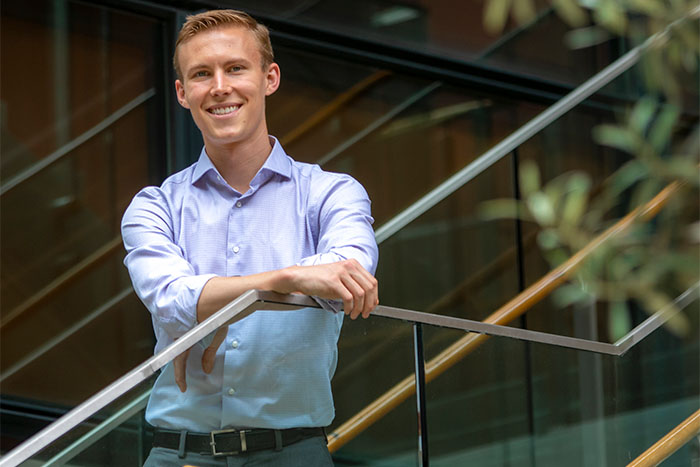
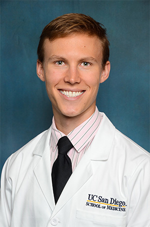
Gregory Sepich-Poore, PhD, entrepreneur, scientist and MD/PhD student at UC San Diego School of Medicine, was recently named to the 2023 Forbes 30 Under 30 List in Healthcare for creating a microbiome-based test for early cancer diagnosis. Additionally, his company, Micronoma, received FDA Breakthrough Designation status for their pioneering lung cancer test in January 2023. Learn about his inspiration for pursuing a career in cancer research and what’s next for this accomplished medical student.
UC San Diego School of Medicine (SOM): Congratulations on being named to the 2023 Forbes 30 Under 30 list for 2023! How did you become interested in cancer research?
Greg Poore: I grew up loving math and science and decided to study biomedical engineering as an undergraduate at Duke University. However, my studies did not have a clear focus until my grandmother was suddenly diagnosed with stage IV pancreatic cancer during my freshman year. She passed away just 33 days later. The experience hit our family like a wrecking ball and left many unanswered questions, including why her disease was diagnosed so late, even after it had metastasized. I longed to find better ways of detecting cancer earlier, which soon led to an insatiable interest in cancer genomics and bioinformatics.
I studied abroad in Singapore, where I conducted cancer genomics research at Duke-NUS Graduate Medical School under Professors Steve Rozen and Patrick Tan. It was my first knee-deep experience using tumor-derived DNA to better understand the differences between malignant and benign tissues, including how this information could be useful to clinicians. For example, one of the research studies we worked on (Tan et al. 2015 Nature Genetics) showed how a patient who originally was diagnosed with a benign breast mass via pathology had mutations identified during sequencing that were more consistent with a borderline malignant lesion. Because of this new data, the biopsy was re-examined by two expert pathologists and re-classified as no longer benign, changing the clinical course of the patient. Through the experiences at Duke-NUS, I realized the importance and beauty of bench-side research in patient care and decided to study cancer as a physician-scientist.
SOM: You recently completed the PhD portion of your studies at UC San Diego in the Jacobs School of Engineering. Can you tell us about your research there?

I began working in Professor Rob Knight’s lab just prior to starting medical school in Fall 2016. I knew very little about the microbiome at the time, but Prof. Knight was a supportive mentor who was willing to take a risk with my cancer genomics background. As it would turn out, the risk-taking would prove fruitful for both of us.
Our initial goal was to re-examine sequencing data from tumors to see if one could discover microbial DNA or RNA ‘hiding’ in the existing information. It’s important to note that the standard workflow for cancer genomics was to map all the sequencing data to a human reference genome and discard everything else, since ‘everything else’ was presumed to be sequencing artifact or contamination. For us, this meant that we were looking for “gold” (microbial DNA) in what others had considered “trash” (DNA that did not map the human genome).
To do this comprehensively for many cancer types in a rapid manner, we mined The Cancer Genome Atlas (TCGA), a public sequencing database of more than 10,000 patients who had 33 different types of cancer. Profiling this much data, which was approximately 600 terabytes, for microbial DNA and RNA was a massive undertaking that required running a supercomputer for months, with the help of Jenya Kopylova, PhD, a post-doctoral research associate in the Knight lab. It was the computational equivalent of searching for needles among bundles of hay.
What we found was surprising and remarkable. Not only were microbes detected in each cancer type, but you could distinguish which sample belonged to which cancer type just using the microbial DNA or RNA. Moreover, trace amounts of the microbial DNA were detectable in matched blood samples in TCGA, meaning you could discern which cancer type a patient had using blood-derived microbial information. We then spent months trying to challenge the results but kept reaching similar conclusions: the microbiomes were cancer type specific.
Eventually, we conducted a study to examine new plasma samples from 100 patients with three types of cancer – lung, prostate, melanoma – and 69 healthy individuals to see if cell-free microbial DNA in plasma could diagnose the presence and type of cancer. Once again, even with dozens of contamination controls, we found that blood-derived microbial DNA was a minimally-invasive, novel biomarker for the presence and type of cancer, leading to a publication in Nature (Poore, Kopylova et al. 2020).
Another independent study from the Weizmann Institute in Israel was published a few months later in Science, making the same conclusions (Nejman et al. 2020). Suddenly, there was a lot of interest in this “cancer microbiome” and how it could be used to improve clinical care. Partnering with that group at the Weizmann among others, we collaborated to write a consensus review on the topic through both historical and scientific lenses (Sepich-Poore et al. 2021 Science), showing how microbes could systemically improve cancer diagnosis, prognosis and therapy.
Towards the end of my PhD, the Weizmann group and ours serendipitously discovered that we were independently working to detect fungi in the cancer cohorts we originally published in 2020. In both cases, fungi replicated the bacterial story: present in dozens of cancer types, distinct between cancer types, and detectable in plasma. The fungi were just much less abundant than their bacterial counterparts. We then combined efforts and analyses to produce the most comprehensive survey of cancer fungi—the “cancer mycobiome”—that was later published as a collaborative paper in Cell (Narunsky-Haziza, Sepich-Poore, Livyatan et al. 2022). Collectively, these papers indicated the non-sterile nature of tumors and the diagnostic opportunities stemming from their microbial residents.
SOM: This is fascinating work. How did this research lead to the establishment of your start-up, Micronoma?
GP: In 2019 I co-founded Micronoma, which is a combination of the words “microbe” and “carcinoma,” alongside two mentors at UC San Diego, Sandrine Miller-Montgomery (former Center for Microbiome Innovation director) and Prof. Knight. The goal of the company is to create microbiome-augmented diagnostics that detect cancer in its earliest stages, so that, personally speaking, people to do not have to go through what my grandmother experienced. More philosophically speaking, I am also a firm believer that entrepreneurship is a necessary vehicle to quickly bring new technologies discovered in the lab to patients in the clinic.
Lung cancer is currently the world’s deadliest cancer type. Micronoma has thus focused on building a novel, early-stage lung cancer diagnostic, OncobiotaLUNG, which recently received FDA Breakthrough Designation status, and we hope to launch the test later this year. We are also actively evaluating the utility of microbial information in other cancer types, including pancreatic, breast, colorectal, and liver cancers. All this work would not have been possible without our amazing 13 team members and the generous financial support from our investors. To date, we have raised $17.5 million.
SOM: Congratulations on these incredible achievements. What’s next on the horizon for you?
GP: We are working very hard on getting Micronoma’s lung cancer test ready for launch later this year, as well as a concomitant paper containing the data that led to the FDA Breakthrough Designation. Career-wise, I am also excited to restart my third year of medical school this summer and remain grateful for the mentors who have supported the completion of my clinical training. It is special to consider that a diagnostic test we developed may soon be prescribable for the patients I serve.
– Stephanie Healey