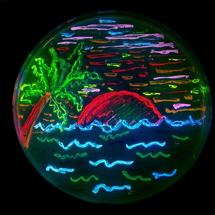
A Petri dish glows whimsically with multi-color fluorescent proteins developed by Roger Tsien. Tsien lab
In 2008, Roger Tsien, who died in 2016, shared the Nobel Prize in chemistry for developing a rich palette of glowing proteins capable of revealing the previously unseen inner workings of cells and organisms.
Imaging technologies based upon green fluorescent protein, derived from a bioluminescent jellyfish, have since revolutionized many aspects of science and medicine, from basic biology to how surgeons operate to identify malignant tissues while avoiding delicate nerves.
Tsien’s profound contributions to science have long been celebrated. Recognition continues. On January 6, he was inducted into the National Inventors Hall of Fame. Here’s a look at some of the research at UC San Diego that has benefitted from Tsien’s illuminating work:
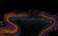
 gfp-mouse retina butterly
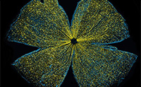
Researchers at UC San Diego Shiley Eye Institute are looking for genetic markers that will help identify age-associated and anatomical aging in eyes. This confocal microscope image depicts a mouse retina sparkling with tell-tale fluorescent molecules.
 gfp-tuszynski
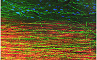
Led by Mark Tuszynski, researchers at UC San Diego have explored using grafted stem cells to repair spinal cord injuries. This image depicts extension of human axons into host adult rat white matter and gray matter three months after spinal cord injury and transplantation of human induced pluripotent stem cell-derived neurons. Green fluorescent protein identifies human graft-derived axons, myelin (red) indicates host rat spinal cord white matter and blue marks host rat gray matter.
 gfp-mouse intestine deerinck
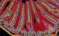
Thomas Deerinck at the National Center for Microscopy and Imaging Research at UC San Diego used multiphoton fluorescence microscopy to capture the nooks and crannies of a section of mouse intestine, with different fluorescent markers indicating different cellular constituents. Actin, a protein that forms the contractile filaments of muscle cells, is shown in green. Lamin, in green, are fibrous proteins that provide structural function. Cell nuclei are depicted in blue.
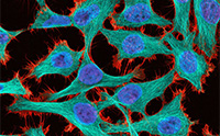 gfp-hela cells-deerinck
In another multiphoton fluorescence image by Deerinck, HeLa cells (a type of immortal cancer cell line used in research) are stained with luminescent markers: red for actin binding toxin phalloidin, cyan for microtubules and blue-purple for cell nuclei.
 gfp-autism gaps-courchesne
Using fluorescent markers to identify different cell types, UC San Diego researcher Eric Courchesne and colleagues showed how patches of disrupted development in the brains of children with autism began during pregnancy.
Watch a 2013 video lecture of Tsien explaining fluorescent proteins and their scientific applications.
— Scott LaFee