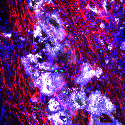
Above: Confocal image of a living tissue slice derived from a patient tumor with metastatic appendiceal cancer (mucinous carcinomatosis peritonei). Immunofluorescence reveals the presence of cancer cells (white), fibroblasts (red) and immune cells (green). Cell nuclei are labeled blue. Photo credit by Jonathan Weitz, UC San Diego Health
Appendiceal cancer is very rare, occurring in perhaps one or two people per 1 million per year. Prognoses is mixed, with a 5-year survival rate of 67 to 97 percent for low-grade tumors detected early, but much lower for advanced cases that may have spread to other parts of the body.
For researchers and physicians, the biggest challenge to studying and treating appendiceal cancer is the lack of an effective preclinical model, something upon which to test new drugs and approaches.
In a recent paper, researchers at UC San Diego School of Medicine and Moores Cancer Center at UC San Diego Health describe creation of the first preclinical model of appendiceal cancer that contains all elements of the tumor, allowing previously stymied investigations to proceed.
“We’ve learned that appendiceal cancer has a distinctive genomic landscape and is surprisingly full of immune cells,” said senior author Andrew Lowy, MD, chief of the Division of Surgical Oncology at Moores Cancer Center at UC San Diego Health and a professor of surgery at UC San Diego School of Medicine.
”Relying on other models, such as colorectal, don’t apply, which makes this an unmet need. Epithelial neoplasms (new, abnormal tissue growth) of the appendix are rare, but without an effective way to study them, the opportunities to develop new treatments have also been rare.”
— Scott LaFee