Image courtesy of Flavio Dell’Acqua, Wellcome Collection.
Since its development in the 1970s and 1980s, magnetic resonance imaging or MRI has transformed both science and medicine, providing a non-invasive tool to peer inside the human body without employing the damaging radiation used in computerized tomography (CT) and positron emission tomography.
MRIs allow scientists and physicians to create two- and three-dimensional representations of virtually every component of the body, including those unwelcome, such as tumors or defects. They are broadly used to assess health and disease.
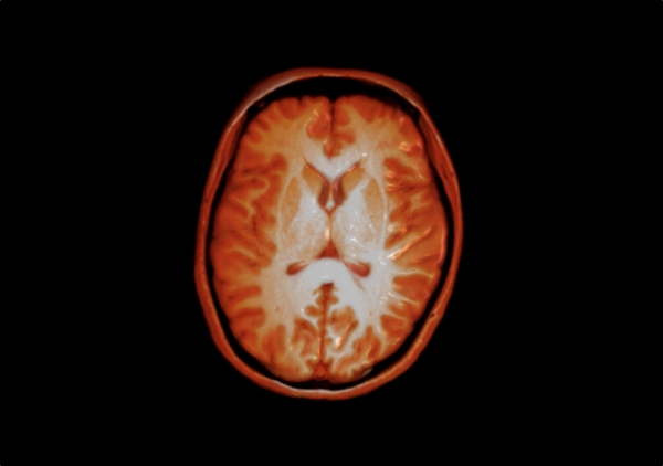
But most MRIs are, by their nature, less than life-like, the product of the body’s natural magnetic properties exploited to create a compilation of scans and a bigger picture. The image above is something different: An MRI slice of a healthy adult brain (viewed from above, front of brain facing top) that has been processes using advanced 3D rendering techniques to mimic the appearance of the brain as it might look after an actual post-mortem dissection. (The orange is a false-color.)
The technique is another way for scientists and physicians can probe what they cannot see, and perhaps see something new.
— Scott LaFee